A raised growth that catches on clothing, a rough patch that makes makeup sit unevenly, or a spot that has gradually become more noticeable can feel like a small concern with an outsized impact on confidence. A benign lesion removal procedure may improve comfort and appearance, but the first step is never simply choosing a treatment. It is confirming that the lesion is appropriate for cosmetic removal and selecting an approach that respects both skin health and the final cosmetic result.
At Natural Beauty Clinic, we believe thoughtful assessment is the foundation of refined, natural-looking care. The goal is not to treat every mark on the skin, but to understand what it is, why it is bothersome, and whether removal is medically appropriate for you.
What is considered a benign skin lesion?
A lesion is a broad medical term for an area of skin that looks or feels different from the surrounding tissue. “Benign” means noncancerous. Common examples can include skin tags, seborrheic keratoses, cherry angiomas, milia, and certain stable moles or other superficial growths.
These lesions are often harmless, although they may itch, become irritated by shaving or jewelry, bleed after friction, or create an unwanted texture or color difference. Some are related to genetics, age, sun exposure, hormonal changes, or simple skin biology. Their presence does not necessarily indicate a problem with overall health.
That said, visual similarity can be misleading. A spot that appears routine may require closer evaluation, and a lesion should not be assumed benign based on an online image or a self-assessment. A careful clinical review is particularly important before any cosmetic procedure that removes or alters tissue.
Why assessment comes before a benign lesion removal procedure
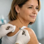
The most important part of a benign lesion removal procedure is often the consultation. We review the lesion’s appearance, location, duration, symptoms, and whether it has changed. Your medical history, medications, history of abnormal scarring, previous skin cancers, immune status, and skin tone can also influence the safest recommendation.
A lesion may need assessment by a physician or referral for dermatologic evaluation if there is uncertainty about its diagnosis. In some cases, a biopsy and pathology review may be appropriate before or during removal. This is especially relevant when a spot is new, changing, irregularly pigmented, persistently crusted, or bleeding without a clear reason.
Patients are sometimes disappointed to learn that a lesion is not suitable for same-day cosmetic treatment. However, that caution is part of responsible care. Removing a suspicious lesion with a cosmetic technique may make later diagnosis more difficult or fail to provide tissue for laboratory assessment when it is needed.
Changes that deserve timely medical attention
Arrange a medical evaluation rather than pursuing cosmetic removal first if a spot is changing in size, shape, color, or sensation. New persistent bleeding, ulceration, pain, itching, or a wound that does not heal should also be reviewed. These signs do not automatically mean a serious condition, but they warrant clinical judgment.
Treatment options depend on the lesion and location
Once a lesion has been assessed as appropriate for removal, the ideal technique depends on its depth, size, type, blood supply, and placement. Skin on the eyelids, lips, nose, chest, and shoulders, for example, may heal differently than skin on the torso or limbs. The trade-off is not simply between “removal” and “no removal.” It is between various methods, healing times, and possible textural or pigment changes.
Shave removal or surgical excision
A superficial raised lesion may sometimes be removed with a shave technique, which levels the growth with the skin’s surface. A deeper lesion or one requiring diagnostic tissue may need surgical excision, where the lesion is removed more completely and the skin may be closed with sutures.
Excision can offer a more definitive approach for selected lesions, but it generally involves a linear scar and a longer healing process. Shave removal may leave less of a line but can still result in temporary redness, altered pigment, or a shallow change in texture. The best option depends on anatomy and clinical findings, not just the smallest possible scar in the short term.
Electrosurgery, curettage, or cryotherapy
Some superficial benign lesions may be treated with carefully controlled heat, scraping, or freezing. These approaches can be useful for certain skin tags, seborrheic keratoses, and other surface growths when clinically appropriate.
With cryotherapy, the treated area may blister, darken, or form a crust before healing. Electrosurgical approaches may create a small scab and temporary redness. Both can carry a risk of lightening or darkening of the skin, particularly in patients prone to post-inflammatory hyperpigmentation. This does not mean these techniques are unsuitable, but it makes pre-treatment planning and aftercare especially important.
Laser-based approaches
Certain benign superficial lesions may be considered for laser treatment after appropriate assessment. Advanced laser technology can be precise, but it is not automatically the preferred option for every growth. Lesion type, skin tone, prior pigmentation concerns, and the need for pathology all matter.
For some patients, laser treatment may support a smooth cosmetic result; for others, another method may be more appropriate or a dermatology referral may be advised. Sophisticated technology is most valuable when it is paired with sound clinical judgment.
What to expect during treatment
Many minor removal procedures are performed in an office setting. Depending on the technique and lesion location, a topical or local anesthetic may be used to support comfort. The treatment itself can be brief, although consultation, preparation, consent, and post-care instructions are equally important parts of the appointment.
Immediately afterward, the site may look pink, red, swollen, or lightly crusted. A small amount of tenderness is common. If a lesion has been excised, you may have sutures and a planned follow-up for removal or wound review. If tissue is sent for pathology, the next steps are guided by the laboratory result and your clinician’s recommendations.
Timing matters when planning treatment. If you have an upcoming vacation, professional event, or significant sun exposure ahead, it may be wise to schedule with enough recovery time. Even when healing progresses smoothly, redness and pigment changes can remain visible longer than patients expect.
Healing well and minimizing visible marks
No procedure can promise scar-free healing. Skin repair is influenced by the removal method, lesion depth, location, genetics, infection risk, sun exposure, and how closely aftercare guidance is followed. The aim is to create the most favorable conditions for healing, while being honest that individual responses vary.
Keep the area clean and follow the wound-care plan provided by your clinician. Avoid picking, rubbing, exfoliating, or applying unapproved active skincare products to a healing site. A crust is part of the normal repair process for many treatments; removing it prematurely can increase irritation and the chance of scarring.
Sun protection is particularly important after lesion removal. Freshly healed skin is more vulnerable to post-inflammatory pigment changes, and ultraviolet exposure can prolong redness or darkening. Once the skin is fully closed and your clinician confirms it is appropriate, scar-support strategies may be considered based on the procedure and your skin’s healing pattern.
Contact your treating clinician promptly if you experience increasing pain, spreading redness, warmth, pus-like drainage, fever, or bleeding that does not stop with gentle pressure. These symptoms do not always indicate a complication, but they should be assessed rather than managed through guesswork.
A personalized approach is worth the extra step
A lesion may be medically benign and still deserve careful planning. The skin is not a blank canvas, particularly in visible areas where subtle differences in texture, pigment, and scar placement matter. Patients with a history of keloids, darker skin tones, melasma, rosacea, or prolonged pigmentation after acne or injury may benefit from a more conservative and individualized approach.
An integrative perspective can also be useful when considering skin quality more broadly. Hydration, nutrition, inflammation, hormones, sleep, sun exposure, and a consistent skincare routine can influence how the skin looks and recovers, although they do not replace appropriate medical assessment or procedural care. When treatment is suitable, our focus is on supporting healthy healing and results that look harmonious with your natural skin.
If a lesion is irritating, changing, or simply affecting how you feel in your skin, begin with an informed evaluation. The most elegant result starts with knowing when removal is appropriate, when pathology or referral is wiser, and which treatment plan best respects your health, comfort, and long-term skin quality.